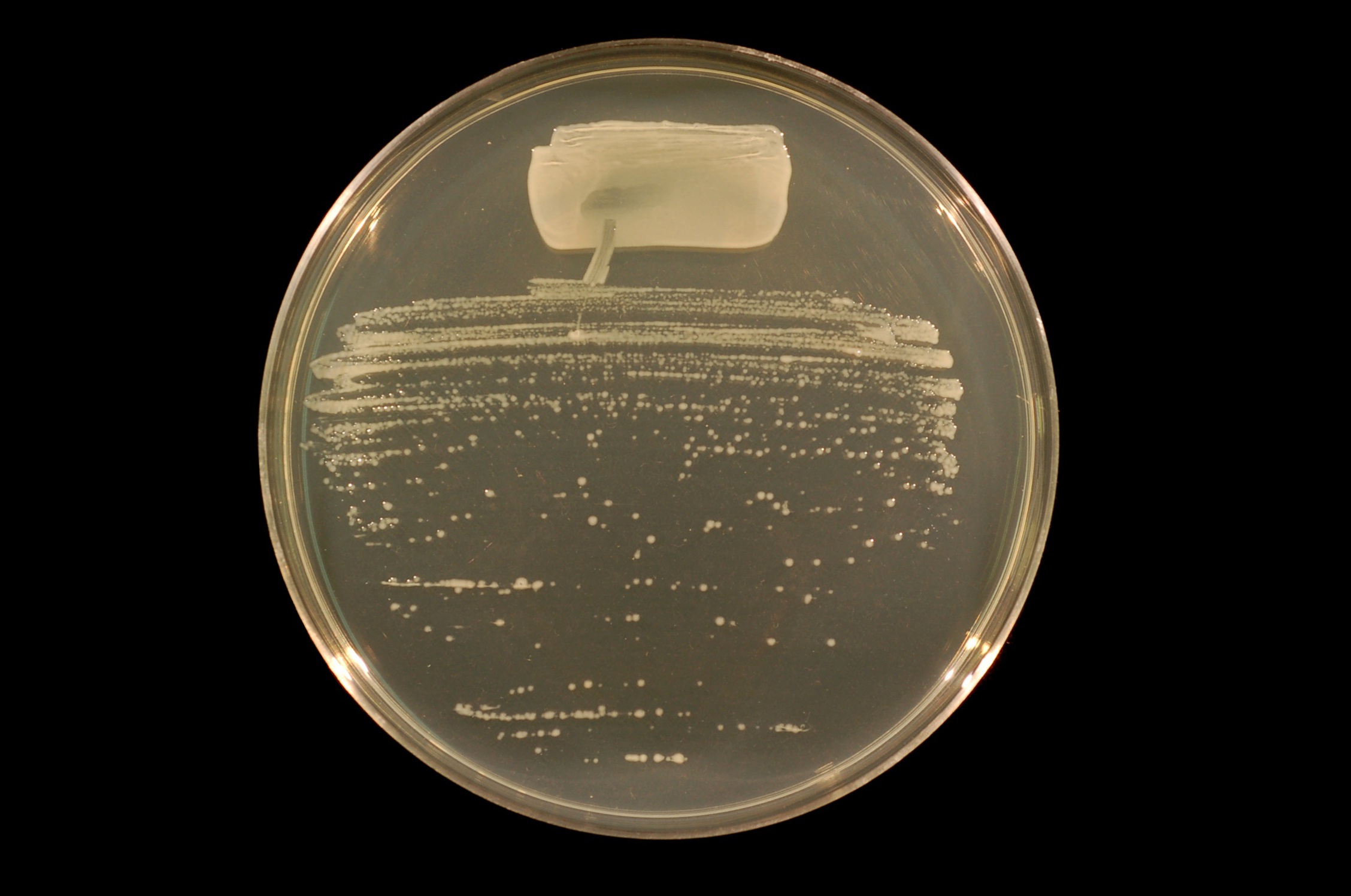
Last year I noticed that there is a paucity of information about nitrogen-fixing rhizobia [1] on Wikipedia. Since I had a lot of rhizobial strains available to me, I decided to photograph them and upload the photographs to Wikimedia Commons. As with the Siberian peashrub photographs [2], I wanted to clean up the background before actually submitting them. I also wanted to center them and rotate them so they didn't look crooked. They sat on the desktop of my lab computer until February of this year. Then I moved them to my jump drive and procrastinated again until now. This week when I had a little free time between experiments at work, I used the GIMP to touch them up.[3] Without further ado, here they are.

Bradyrhizobium japonicum forms a symbiosis on the roots of soybean (Glycine max).

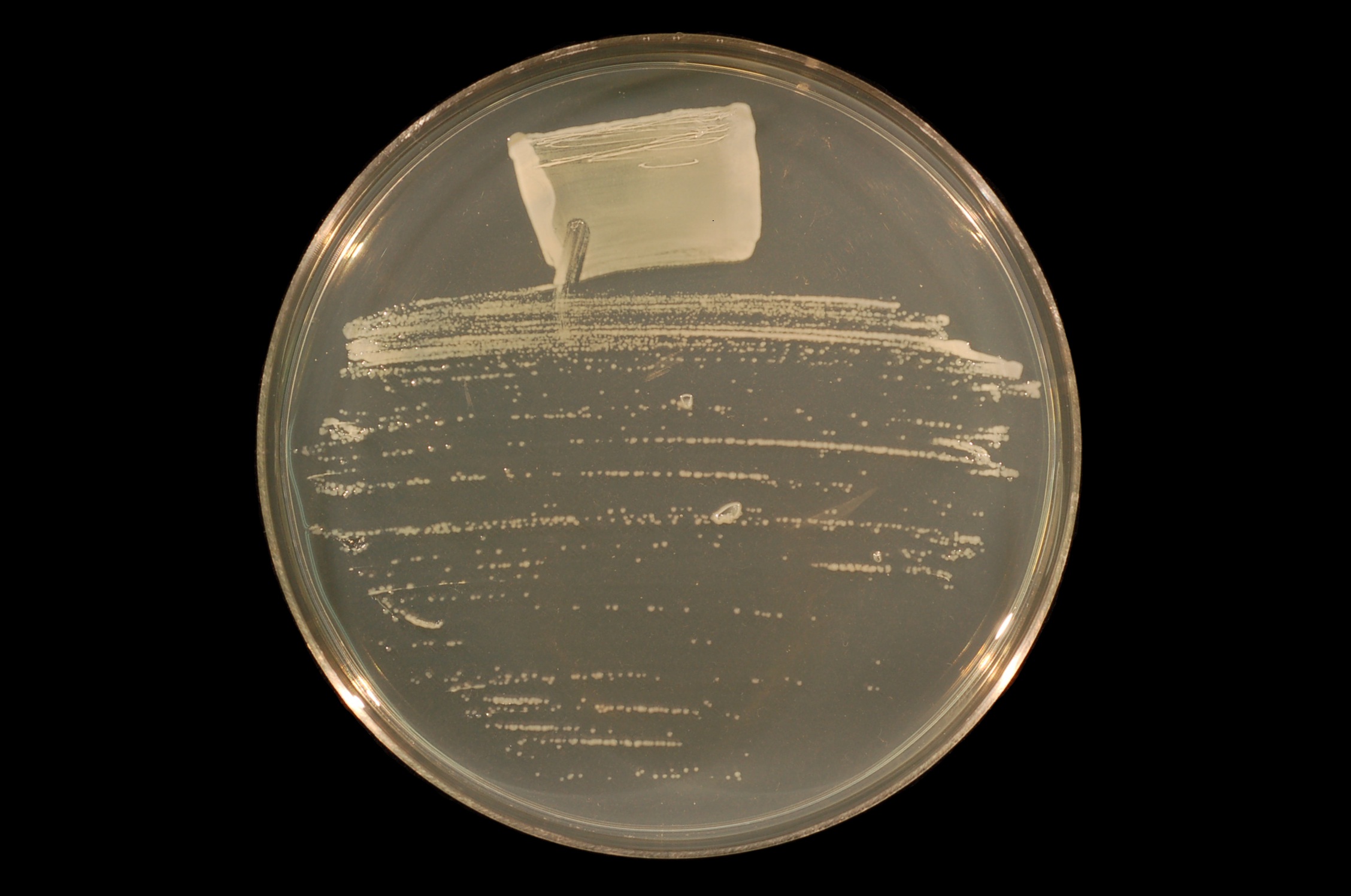
Mesorhizobium loti forms a symbiosis on the roots of deervetch (Lotus japonicus).
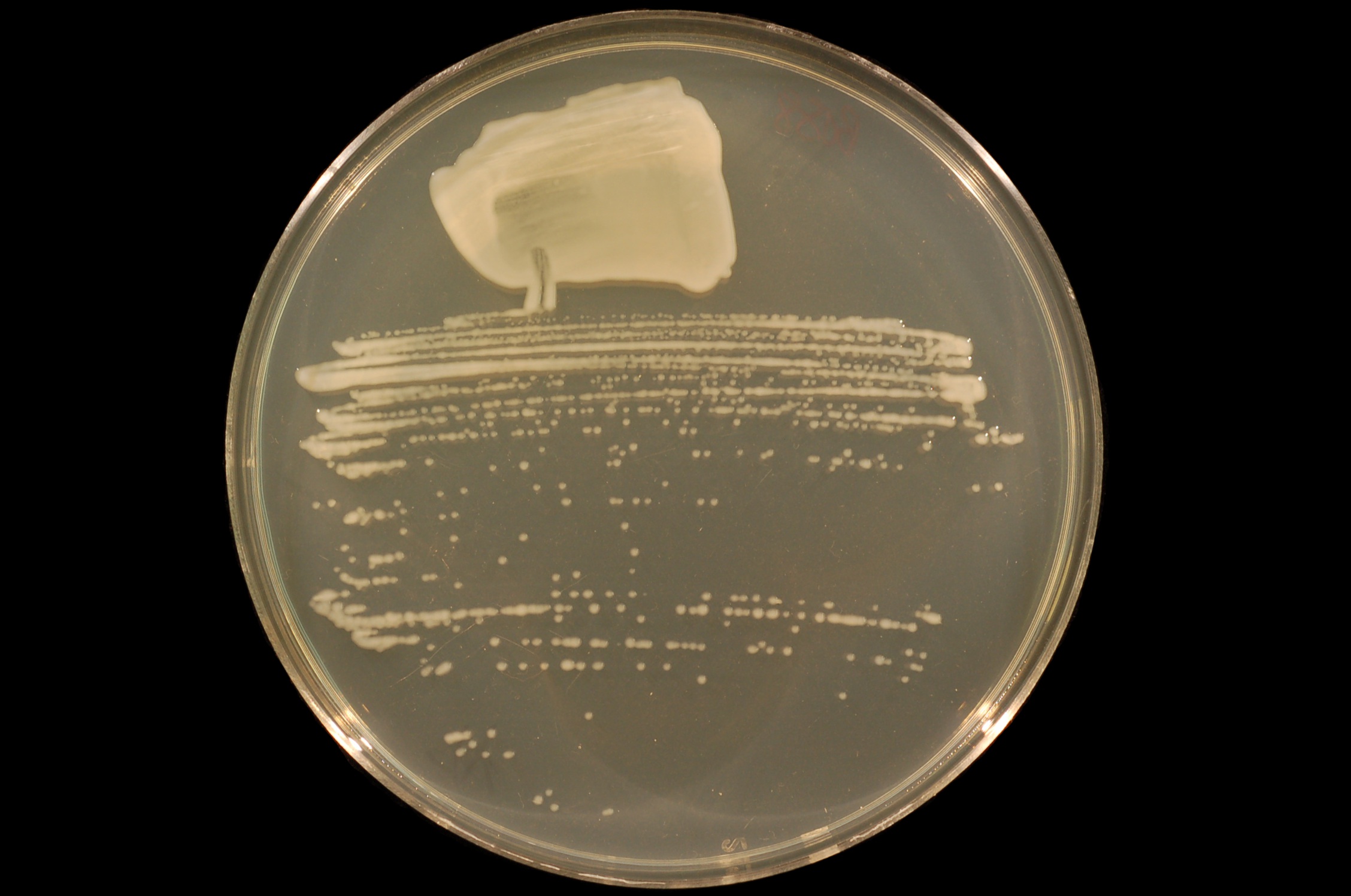
Rhizobium tropici forms a symbiosis with the common bean (Phaseolus vulgaris).
Sinorhizobium fredii forms a symbiosis with soybean (Glycine max).

Sinorhizobium medicae forms a symbiosis with toothed burclover (Medicago polymorpha).

Sinorhizobium meliloti forms a symbiosis with alfalfa (Medicago sativa). This is the bacteria I do most of my work with.
Sinorhizobium sp. NGR234 [4] forms a symbiosis with over 112 different genera of plants.
Now up to this point, you're probably thinking that they all look the same. And you are not incorrect. But, read on and I'll show you a little more variety.

Bacillus mycoides isn't a rhizobial bacterium. But it is still interesting because when you grow it in a Petri dish, it always grows in spirals. Some strains always spiral clockwise; others always spiral counterclockwise. Scientists still haven't figured out how. This one spirals clockwise.


Pseudomonas fluorescens is one of the bacteria used to produce yoghurt. It isn't rhizobial. When you look at it on a Petri dish it is cream-colored, or may even look pale green. However, when you shine ultraviolet light on it, it will glow a bright cyan (bottom). Many species of Pseudomonas secrete chemicals, known as pyocyanins and pyoverdins, which help them to scavenge iron from their environment. These are what glow under UV light.
Once I had all of these images uploaded, I inserted them into the appropriate (existing) pages on Wikipedia: Bacillus mycoides, Bradyrhizobium japonicum, Mesorhizobium, Pseudomonas fluorescens, Rhizobium, Sinorhizobium, and Sinorhizobium meliloti.
Notes:
[1] If you don't know what nitrogen fixation is or rhizobia, see my post What Is It That Matt Does, Anyway?
[2] See my post The End of Procrastination II.
[3] If you don't know what the GIMP is or what it is used for, see my post Raster Graphics and Vector Graphics.
[4] It's species has never formally been declared, but it is believed to belong to Sinorhizobium fredii.
Image attributions:
Bradyrhizobium japonicum is by Ninjatacoshell, available at http://commons.wikimedia.org/wiki/File:Bradyrhizobium japonicum USDA 110 on TY agar.JPG. The original can be seen here.
Mesorhizobium loti is by Ninjatacoshell, available at http://commons.wikimedia.org/wiki/File:Mesorhizobium loti strain NZP2037 on TY agar.JPG. The original can be seen here.
Rhizobium tropici is by Ninjatacoshell, available at http://commons.wikimedia.org/wiki/File:Rhizobium tropici strain BR816 on TY agar.JPG. The original can be seen here.
Sinorhizobium fredii is by Ninjatacoshell, available at http://commons.wikimedia.org/wiki/File:Sinorhizobium fredii strain USDA257 on TY agar (clean).JPG. The original can be seen here.
Sinorhizobium medicae is by Ninjatacoshell, available at http://commons.wikimedia.org/wiki/File:Sinorhizobium medicae on TY agar.JPG. The original can be seen here.
Sinorhizobium meliloti is by Ninjatacoshell, available at http://commons.wikimedia.org/wiki/File:Sinorhizobium meliloti strain Rm1021 on TY agar.JPG. The original can be seen here.
Sinorhizobium sp. strain NGR234 is by Ninjatacoshell, available at http://commons.wikimedia.org/wiki/File:Sinorhizobium strain NGR234 on TY agar.JPG. The original can be seen here.
Bacillus mycoides is by Ninjatacoshell, available at http://commons.wikimedia.org/wiki/File:Bacillus mycoides on TY agar.JPG. The original can be seen here.
Pseudomonas fluorescens under white light is by Ninjatacoshell, available at http://commons.wikimedia.org/wiki/File:Pseudomonas fluorescens on TY agar (white_light).JPG. The original can be seen here.
Pseudomonas fluorescens under ultraviolet light is by Ninjatacoshell, available at http://commons.wikimedia.org/wiki/File:Pseudomonas fluorescens on TY agar (UV_light).JPG. The original can be seen here.
Bradyrhizobium japonicum forms a symbiosis on the roots of soybean (Glycine max).
Mesorhizobium loti forms a symbiosis on the roots of deervetch (Lotus japonicus).
Rhizobium tropici forms a symbiosis with the common bean (Phaseolus vulgaris).
Sinorhizobium fredii forms a symbiosis with soybean (Glycine max).
Sinorhizobium medicae forms a symbiosis with toothed burclover (Medicago polymorpha).
Sinorhizobium meliloti forms a symbiosis with alfalfa (Medicago sativa). This is the bacteria I do most of my work with.
Sinorhizobium sp. NGR234 [4] forms a symbiosis with over 112 different genera of plants.
Now up to this point, you're probably thinking that they all look the same. And you are not incorrect. But, read on and I'll show you a little more variety.
Bacillus mycoides isn't a rhizobial bacterium. But it is still interesting because when you grow it in a Petri dish, it always grows in spirals. Some strains always spiral clockwise; others always spiral counterclockwise. Scientists still haven't figured out how. This one spirals clockwise.
Pseudomonas fluorescens is one of the bacteria used to produce yoghurt. It isn't rhizobial. When you look at it on a Petri dish it is cream-colored, or may even look pale green. However, when you shine ultraviolet light on it, it will glow a bright cyan (bottom). Many species of Pseudomonas secrete chemicals, known as pyocyanins and pyoverdins, which help them to scavenge iron from their environment. These are what glow under UV light.
Once I had all of these images uploaded, I inserted them into the appropriate (existing) pages on Wikipedia: Bacillus mycoides, Bradyrhizobium japonicum, Mesorhizobium, Pseudomonas fluorescens, Rhizobium, Sinorhizobium, and Sinorhizobium meliloti.
Notes:
[1] If you don't know what nitrogen fixation is or rhizobia, see my post What Is It That Matt Does, Anyway?
[2] See my post The End of Procrastination II.
[3] If you don't know what the GIMP is or what it is used for, see my post Raster Graphics and Vector Graphics.
[4] It's species has never formally been declared, but it is believed to belong to Sinorhizobium fredii.
Image attributions:
Bradyrhizobium japonicum is by Ninjatacoshell, available at http://commons.wikimedia.org/wiki/File:Bradyrhizobium japonicum USDA 110 on TY agar.JPG. The original can be seen here.
Mesorhizobium loti is by Ninjatacoshell, available at http://commons.wikimedia.org/wiki/File:Mesorhizobium loti strain NZP2037 on TY agar.JPG. The original can be seen here.
Rhizobium tropici is by Ninjatacoshell, available at http://commons.wikimedia.org/wiki/File:Rhizobium tropici strain BR816 on TY agar.JPG. The original can be seen here.
Sinorhizobium fredii is by Ninjatacoshell, available at http://commons.wikimedia.org/wiki/File:Sinorhizobium fredii strain USDA257 on TY agar (clean).JPG. The original can be seen here.
Sinorhizobium medicae is by Ninjatacoshell, available at http://commons.wikimedia.org/wiki/File:Sinorhizobium medicae on TY agar.JPG. The original can be seen here.
Sinorhizobium meliloti is by Ninjatacoshell, available at http://commons.wikimedia.org/wiki/File:Sinorhizobium meliloti strain Rm1021 on TY agar.JPG. The original can be seen here.
Sinorhizobium sp. strain NGR234 is by Ninjatacoshell, available at http://commons.wikimedia.org/wiki/File:Sinorhizobium strain NGR234 on TY agar.JPG. The original can be seen here.
Bacillus mycoides is by Ninjatacoshell, available at http://commons.wikimedia.org/wiki/File:Bacillus mycoides on TY agar.JPG. The original can be seen here.
Pseudomonas fluorescens under white light is by Ninjatacoshell, available at http://commons.wikimedia.org/wiki/File:Pseudomonas fluorescens on TY agar (white_light).JPG. The original can be seen here.
Pseudomonas fluorescens under ultraviolet light is by Ninjatacoshell, available at http://commons.wikimedia.org/wiki/File:Pseudomonas fluorescens on TY agar (UV_light).JPG. The original can be seen here.
You probably intended "secrete" instead of "secret" in the penultimate paragraph. Nice pics, but the first ones did look alike.
ReplyDeleteFixed. Thanks.
ReplyDelete